
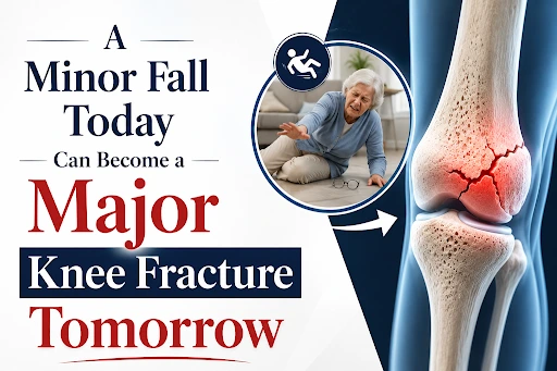
A minor slip should not cause an orthopaedic emergency. Yet, for many residents, a simple misstep frequently results in a complex, life-altering knee fracture due to underlying osteoporosis.
This “silent disease” erodes bone strength from the inside out, making weight-bearing joints highly fragile.
If you or an elderly family member is facing intense pain or an inability to walk after a minor fall, seek immediate care from an expert knee fracture doctor in Jamshedpur, like Dr Prashant Arya, to protect your long-term mobility.
Quick Answer: Can Osteoporosis Cause Knee Fractures?
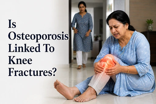
- Yes. Osteoporosis severely degrades internal trabecular bone scaffolding, turning minor household slips into complex joint fractures.
- Common Break Sites: Structural bone failure typically occurs at the distal femur (lower thigh), tibial plateau (upper shin), or the patella (kneecap).
- Critical Warning Signs: Seek emergency medical evaluation if a fall results in an inability to bear weight, rapid swelling, or visible limb deformity.
- Clinical Solution: Consult an experienced knee fracture doctor in Jamshedpur for definitive digital imaging (X-ray/CT/DEXA) and advanced fixed-angle surgical fixation to safely restore long-term mobility.
Understanding Osteoporosis and Bone Fragility
What Happens to Bones in Osteoporosis?
Osteoporosis is a progressive metabolic bone disease in which bone resorption outpaces new bone formation. This structural imbalance strips essential minerals, such as calcium, from the skeletal matrix, transforming a healthy, dense framework into a fragile, porous one.
Consequently, the bone loses its internal structural reinforcement, rendering it incapable of safely resisting everyday mechanical forces and weight-bearing loads.
Common Risk Factors for Osteoporosis
- Advanced Age: A natural decline in the body’s bone remodelling cycles over time.
- Hormonal Depletion: Accelerated bone mineral loss is commonly observed in postmenopausal women due to dropping estrogen levels.
- Nutritional Deficiencies: A chronic, systemic lack of adequate dietary Calcium and active Vitamin D3 tracking.
- Lifestyle Factors: Prolonged physical inactivity, desk-bound routines, smoking, or excessive alcohol intake.
Healthy Bone vs Osteoporotic Bone
Healthy bone contains a thick, tightly interwoven scaffolding of trabecular (spongy) tissue designed to absorb and distribute mechanical impacts. In contrast, osteoporotic bone features thin, brittle cross-links with widened internal gaps.
This compromised structure has minimal mechanical elasticity, making it highly prone to snapping under minor stress or sudden torsional loading.
Why Osteoporosis Makes Knee Fractures More Likely
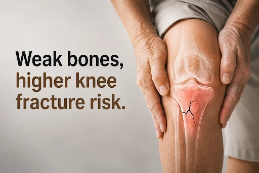
The Role of Trabecular Bone Around the Knee
Osteoporosis targets the internal spongy (trabecular) bone structure, which directly compromises the knee joint’s weight-bearing foundation.
- Load Dynamics: The knee acts as a primary skeletal pillar, absorbing forces up to four times your total body weight during routine movement.
- Structural Thinning: The expanded ends of the femur and tibia rely on a dense trabecular network to cushion these heavy physical loads.
- Systemic Collapse: Metabolic bone thinning rapidly strips away these internal cross-links, leaving the joint base structurally hollow.
Why Low-Energy Falls Become Dangerous
Brittle, osteoporotic bone lacks the mechanical elasticity needed to absorb routine everyday impacts.
- Impact Failure: A simple trip on a flat floor or a slip in the kitchen bypasses the normal muscular shock-absorbing mechanism.
- Force Overload: Low-energy impacts deliver sudden kinetic forces that instantly exceed the ultimate breaking strength of thin bone.
- Severe Outcomes: Minor household bumps that would leave healthy bone uninjured result in complex, multi-fragment joint fractures.
What Is a Fragility Fracture?
A fragility fracture is clinically defined as any bone break resulting from an impact equal to or less than a simple fall from standing height.
- Diagnostic Marker: Sustaining a fracture from a minor household fall confirms severe, unmanaged underlying bone loss.
- Clinical Indicator: It serves as a definitive warning sign that systemic bone mineral density has dropped to dangerously low levels.
- Urgent Care Protocol: This injury requires immediate, dual-action medical intervention combining trauma surgery with aggressive osteoporosis therapies.
Which Knee Fractures Are Common in Osteoporosis Patients?
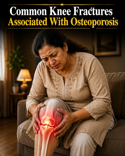
Distal Femur Fracture
This fracture involves a break occurring at the lower segment of the thigh bone, just above the main knee joint line.
- Symptoms: Severe localised pain above the kneecap, rapid joint swelling, and a complete inability to lift the leg.
- Potential Complications: High risk of hardware failure, such as screw pull-out, if standard fixation is anchored into soft bone.
Tibial Plateau Fracture
This injury compromises the flat upper surface of the shin bone, which directly supports the joint’s cartilage cushion.
- Symptoms: Intense pain aggravated by weight-bearing and severe bruising around the top segment of the shin.
- Impact on Knee Stability: Leads to immediate structural collapse, creating a high risk of chronic post-traumatic arthritis if left misaligned.
Patellar Fracture
A patellar fracture involves a clean break or shattering of the kneecap shield sitting directly over the front of the joint.
- Symptoms: Distinct localised tenderness directly over the kneecap and a palpable physical gap in the bone matrix.
- Functional Limitations: Completely disrupts the leg’s extensor mechanism, preventing patients from straightening their limb independently.
Comparison Table of Osteoporotic Knee Fractures
| Fracture Classification | Primary Bone Involved | Common Injury Mechanism | Initial Impact on Joint Stability |
| Distal Femur | Thigh Bone | Direct impact on the thigh or landing hard on an extended leg. | Severe instability; it completely prevents straight-leg lifting. |
| Patellar | Kneecap | Direct forward fall landing squarely on the kneecap. | Loss of the knee extension mechanism; high localised swelling. |
| Tibial Plateau | Shin Bone | Minor twisting fall or sudden vertical loading step. | Complete inability to bear weight; high risk of joint misalignment. |
Warning Signs and Diagnosis Protocols For Knee Fractures
Symptoms Requiring Immediate Medical Attention
If you or an elderly relative experiences a fall inside your home, carefully monitor the joint for these acute symptoms:
- Severe Pain: Intense, localised agony that does not subside with rest.
- Swelling: Rapid fluid or blood accumulation filling the joint space.
- Inability to Bear Weight: Failure to stand or walk even two steps.
- Reduced Movement: Complete inability to flex or extend the limb.
- Instability: A distressing sensation that the joint is giving way.
- Deformity: A visible structural shift where the leg appears crooked.
How a Knee Fracture Doctor Diagnoses Brittle Bone Injuries
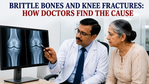
Clinical Patellofemoral & Tibiofemoral Evaluation
- Integumentary check: Assesses for open fractures, soft-tissue envelope compromise, and hemarthrosis.
- Palpation localised: Isolates joint line tenderness and focal exoskeletal crepitus.
- Kinesiology mapping: Measures passive range of motion to detect mechanical blocks.
- Neurovascular status: Audits distal pulses and peroneal/tibial nerve conduction.
- Comorbidity review: Flags osteopenia markers, age-related osteogenesis lag, and metabolic risks.
Digital Orthogonal Radiography
- Frontline triage: Employs anteroposterior (AP) and lateral X-rays for baseline screening.
- Structural displacement: Quantifies cortical disruption, trabecular shearing, and axial malalignment.
High-Resolution Computed Tomography (CT)
- Volumetric reconstruction: Generates 3D cross-sectional slices of comminuted fractures.
- Pre-operative planning: Maps intra-articular extension for optimising the open reduction internal fixation (ORIF) trajectory.
Magnetic Resonance Imaging (MRI)
- Occult detection: Identifies subchondral micro-fractures and bone marrow oedema.
- Ligamentous integrity: Diagnoses concomitant tears in the cruciate (ACL/PCL), collateral (MCL/LCL) ligaments, and menisci.
Dual-Energy X-Ray Absorptiometry (DEXA)
- Densitometry profiling: Calculates central skeletal T-scores to diagnose structural porosity.
- Secondary prevention: Informs antiresorptive therapy or anabolic bone-building protocols post-surgery.
Treatment Options for Osteoporotic Knee Fractures
 Non-Surgical Pathways
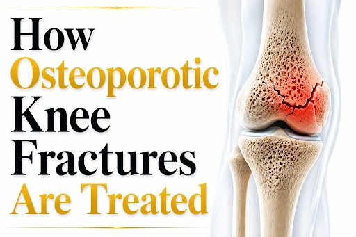 Non-Surgical Pathways
- Bracing & Immobilisation: Using an adjustable long-leg rigid brace and strict non-weight-bearing walker protocols to allow non-displaced fractures to knit together safely.
- Physiotherapy & Management: Gentle non-weight-bearing isometric exercises paired with medical therapies to stimulate bone mineral deposition while the fracture heals.
Specialised Surgical Treatment
- Locking Plate Fixation & Nailing: Advanced fixed-angle plates lock screws into the plate itself, creating a stable construct that prevents hardware from backing out of soft bone. Intramedullary nails stabilise specific shaft fractures.
- Joint Replacement & Planning: Transitioning to a primary knee replacement if the bone is too brittle to hold screws. Specialised surgeons use advanced techniques, such as injecting biocompatible bone cement around screw threads, to provide a solid foundation within the weakened bone matrix.
Recovery, Prevention & Early Intervention of Knee Fractures
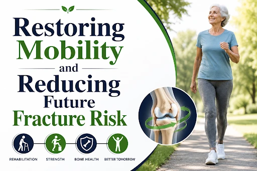
Expected Recovery Timeline & Rehabilitation
The healing journey spans 3 to 6 months. Bone tissue bridging occurs within 8 to 12 weeks, followed by remodelling.
Structured physical therapy is vital to optimal recovery, helping senior folks rebuild weakened quadriceps, regain flexibility, restore balance, and reduce fall risk.
Factors affecting healing include bone density, age, fracture severity, and conditions like diabetes.
Preventing Future Knee Fractures
- Nutritional Support & Strength: Ensure daily Calcium intake paired with therapeutic Vitamin D3 doses to facilitate absorption. Low-impact activities focused on quad strengthening enhance coordination.
- Fall Prevention Strategies: Modify your living environment by removing loose area rugs, installing secure grab bars in the bathroom, and keeping hallways brightly lit.
- Medical Screening & Treatment: Adults over 50 should schedule routine bone density screenings. If a DEXA scan confirms low bone density, specialists prescribe medical therapies—such as bisphosphonates or monoclonal antibodies—to slow bone loss and rebuild bone strength.
Risks of Delayed Treatment
- Malunion & Deformity: The bone heals in an incorrect, crooked position, causing permanent limb deformity.
- Joint Stiffness & Pain: Severe scar tissue formation restricts walking ability and causes chronic pain from cartilage misalignment.
- Loss of Independence: Long-term reliance on walking aids reduces a patient’s ability to perform daily activities independently.
Final Takeaway:
Get your knee injury assessed early by Dr Prashant Arya, MBBS, MS Orthopaedics, a trusted knee fracture doctor in Jamshedpur.
Osteoporotic and fragile bone fractures often require specialised evaluation and advanced diagnostic assessment to prevent complications, restore mobility, and support faster recovery.
Do not ignore the warning signs. Call 091632 83798 for a private consultation today.
People Also Ask
Can osteoporosis cause knee fractures after a minor fall?
Ans: Yes, osteoporosis weakens the internal bone structure so severely that a simple slip can easily fracture the knee. When bone density is low, the joint loses its structural elasticity, causing the proximal tibia or distal femur to crack under low-energy impacts that healthy bone would easily absorb.
What is the most common osteoporotic knee fracture?
Ans: The two most frequent injuries seen in patients with low bone density are distal femur fractures and tibial plateau fractures. These areas are rich in cancellous bone, which thins out rapidly due to osteoporosis, making the regions just above and below the knee joint highly vulnerable to crushing forces during a fall.
Do all knee fractures require surgery?
Ans: No, stable and non-displaced fractures can be treated non-surgically using rigid bracing and strict non-weight-bearing protocols. However, if the bone fragments have shifted or telescoped into each other, surgical intervention using fixed-angle locking compression plates is mandatory to prevent long-term limb malalignment.
How long does recovery take?
Ans: The recovery timeline typically spans 3 to 6 months, depending on fracture severity and the patient’s baseline bone quality. Because osteoporotic bone heals more slowly, a structured, step-by-step physical therapy routine is vital to safely restore joint mobility without risking hardware failure or permanent stiffness.
Can osteoporosis be treated after a fracture?
Ans: Yes, medical management for osteoporosis should be initiated immediately following a fracture to aid healing and reduce future injury risks. Specialists coordinate targeted treatments, which may involve specialised anti-resorptive medications and nutritional support to actively improve systemic bone mineral density during recovery